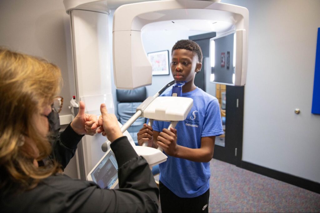
One of the most important elements of our process as an orthodontic office is digital imaging and X-rays. This technology gives us incredible access and precision when it comes to treating our patients, and today, Smiles from the Hart is here to explain further: How are X-rays and imaging used to track progress in orthodontics?
X-Rays and Imaging
There are various imaging methods we can utilize for patients based on their situation.
- Intraoral X-rays: These are the most common types of dental imaging. They involve placing a small sensor of film in the patient’s mouth to capture detailed images of individual teeth, roots, and surrounding bones. They can assess bone levels, detect cavities, and evaluate the health of tooth roots.
- Panoramic X-rays: These provide a broad view of the entire mouth, including all the teeth, upper and lower jaws, and relative structures. We use these for assessing overall dental health, identifying impacted teeth, and planning orthodontic treatment or oral surgery.
- Digital Imaging: These types of technology, such as digital sensors and intraoral cameras, capture high-resolution images of the teeth and gums. These images can be viewed instantly on a computer screen, allowing dentists to visualize dental conditions, educate patients, and plan treatment more effectively.
How We Use Them
With these processes in mind, here are some of the specific ways we utilize them in our treatment plans as orthodontists:
- Initial Diagnosis and Treatment Planning: This imaging is used in the initial diagnosis and treatment planning phase. They provide us with extremely detailed information on the position of the teeth, the relationship between the jaws, the presence of impacted teeth, and any underlying skeletal abnormalities.
- Monitoring Tooth Movement: X-rays and imaging are used to monitor the progress of tooth movement during the course of orthodontic treatment. Serial periapical or panoramic radiographs can be taken at regular intervals to assess the position of the teeth and track their movement over time.
- Evaluation of Root Resorption: We can evaluate the extent of root resorption, which is the shortening of tooth roots that can occur as a result of orthodontic treatment. By periodically assessing root length and morphology, orthodontists can identify any signs of excessive root resorption and adjust treatment as needed to minimize further damage.
- Assessment of Treatment Stability: After orthodontic treatment is completed, X-rays and imaging are both used to evaluate the stability of the treatment outcomes. By comparing post-treatment images to baseline images taken before treatment, Dr. Hart can assess any changes that may have occurred and determine if additional inventions are necessary to maintain the results.
- Identification of Orthodontic Emergencies: We can use X-rays to identify and diagnose orthodontic emergencies, such as loose brackets, broken wires, or displaced teeth. By being able to visualize the underlying structures of the teeth and jaws, X-rays can assist in determining the appropriate course of action to address these issues promptly and effectively.
- Communication with Patients and Referring Providers: Imaging can provide us with visual representations of the patient’s dental and skeletal anatomy, which can be helpful for educating patients and referring providers about the nature of their orthodontic problems and the proposed treatment plan.
FAQs
It’s perfectly normal to have questions about the diagnostic tools we use in our office. Dr. Hart will always be the best resource for these questions, but let’s cover some of them here!
Q: Are dental X-rays safe?
Yes, they’re considered safe when performed with the appropriate equipment and techniques. The amount of radiation exposure from dental X-rays is minimal, and modern X-ray systems further reduce doses compared to traditional film X-rays.
Q: Can pregnant women have dental X-rays taken?
While these are generally safe, pregnant women should always avoid unnecessary exposure to radiation, especially during the first trimester when the fetus is most sensitive to radiation. However, if the X-ray is necessary, we will take appropriate shielding measures and precautions to minimize exposure to the fetus.
Q: How often should I have X-rays taken?
The frequency of your dental X-rays is highly dependent on factors such as your age, dental health, and risk of oral diseases. Generally, X-rays may be recommended at routine dental check-ups every six to twelve months. Patients with a history of dental problems or undergoing specific treatments may require them more frequently.
A Clearer View
Whether you’re new to orthodontics or you’ve had some prior treatment, Dr. Hart and our team are absolutely thrilled to get to help you on this journey. X-rays and imaging are just a couple of the many tools we use to get you the best possible results. If you’re looking into our services, give us a call at our Murfreesboro (615-890-7246) or Gallatin (615-452-2868) offices.


